Trigger Finger: Why Does My Finger Lock or Click?

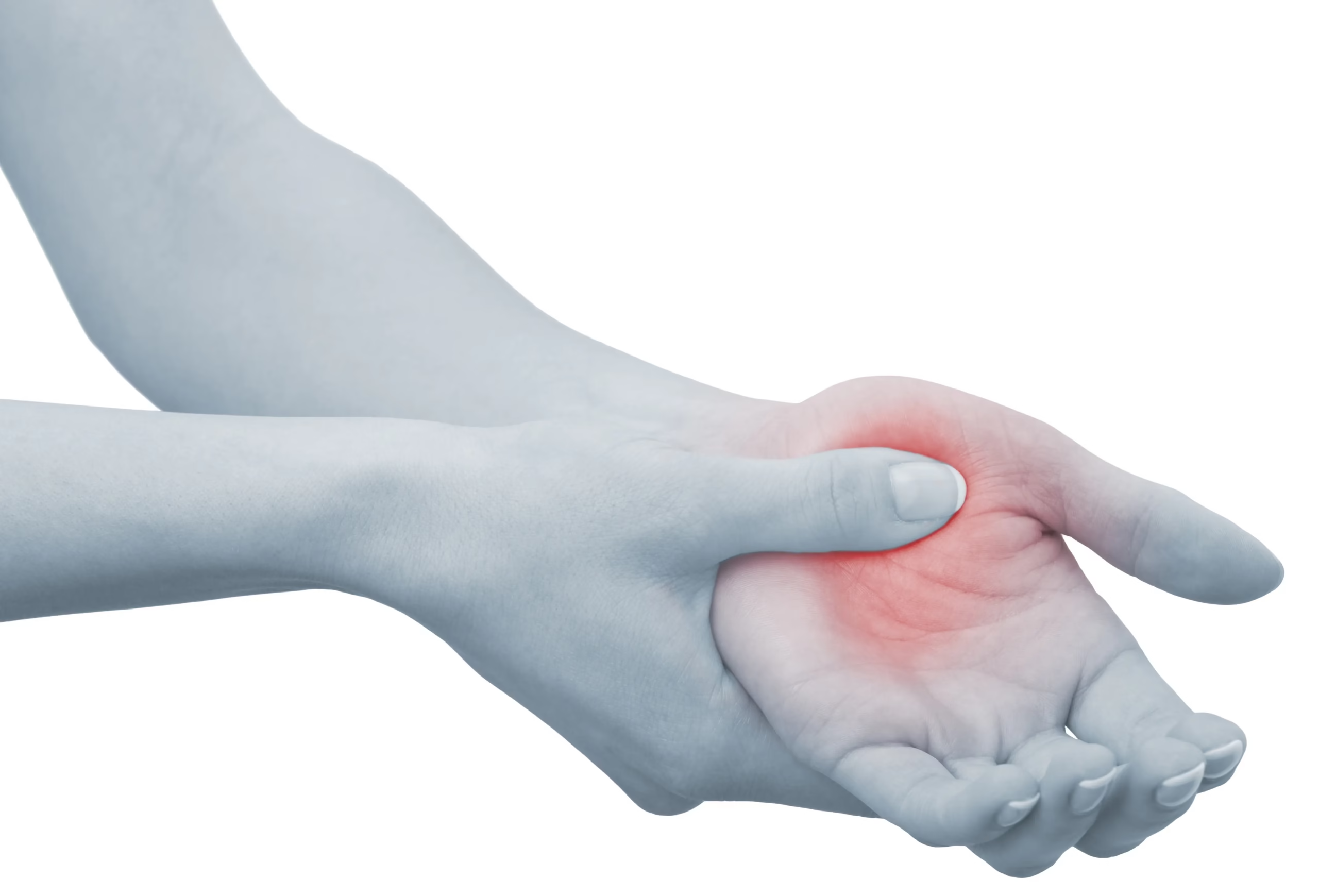
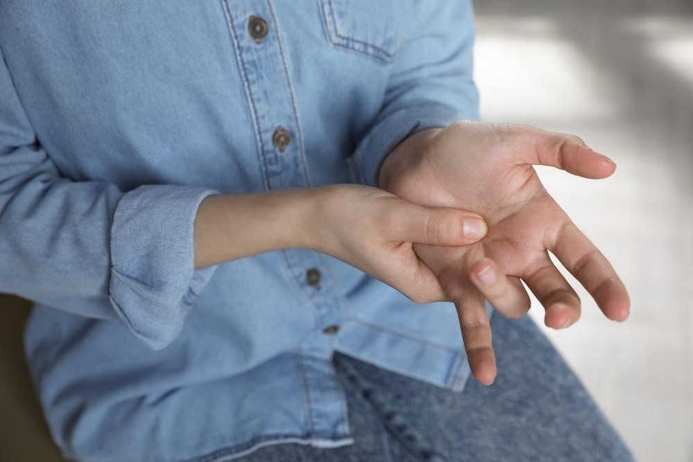
Have you ever noticed your finger catching or locking when you try to straighten it? Perhaps it clicks painfully before suddenly releasing, much like pulling a trigger. This common condition is known as trigger finger, or stenosing tenosynovitis.

Trigger finger can affect anyone, although it is more common in middle-aged adults and people who frequently perform repetitive gripping activities. While symptoms may begin as a minor annoyance, they can progress to painful locking that interferes with everyday tasks.

The good news is that trigger finger is highly treatable. Most patients improve with conservative treatment, and when surgery is necessary, it is typically a simple procedure with excellent outcomes.

What Is Trigger Finger?

The tendons that bend your fingers glide through small tunnels called tendon sheaths. These sheaths are lined with a smooth tissue that allows the tendons to move freely during gripping and finger motion.

At the base of each finger is a structure called the A1 pulley, which keeps the tendon close to the bone. In trigger finger, inflammation or thickening of the tendon or pulley narrows this space. As a result, the tendon no longer glides smoothly and begins to catch during movement.

Initially, this causes clicking. As the condition progresses, the finger may lock in a bent position and require assistance to straighten.

Who Is at Risk?

Trigger finger can develop in anyone but is more common in:

  • Adults between 40 and 60 years of age
  • Women
  • Individuals with diabetes
  • Patients with rheumatoid arthritis
  • People who perform repetitive gripping or forceful hand activities
  • Workers who regularly use vibrating tools

The thumb, ring finger, and middle finger are most commonly affected, although any finger can develop the condition.

Common Symptoms

Symptoms often begin gradually and may include:

  • Pain or tenderness at the base of the finger
  • A clicking or snapping sensation during movement
  • Morning stiffness
  • A feeling that the finger catches when bending
  • A finger that locks in a bent position
  • A small tender lump in the palm near the affected finger

Many patients notice that symptoms are worse in the morning and improve slightly as the hand warms up.

How Is Trigger Finger Diagnosed?

In most cases, trigger finger is diagnosed during a clinical examination.

Your orthopedic surgeon will:

  • Discuss your symptoms
  • Examine finger motion
  • Feel for tenderness over the A1 pulley
  • Assess whether the finger catches or locks
  • Evaluate hand strength and function

Imaging studies such as X-rays are usually unnecessary unless another condition is suspected. Ultrasound may occasionally be used to evaluate the tendon and pulley.

Can Trigger Finger Go Away on Its Own?

In mild cases, symptoms may improve with activity modification and rest. However, untreated trigger finger can become progressively more painful and may eventually lock completely.

Seeking treatment early often leads to faster recovery and reduces the likelihood of surgery.

Conservative Treatment

Activity Modification

Reducing repetitive gripping or forceful hand activities can decrease irritation of the tendon and allow inflammation to settle.

Anti-inflammatory Medication

Short courses of anti-inflammatory medication may help relieve pain during the early stages, although they do not correct the mechanical narrowing responsible for the condition.

Splinting

A finger splint worn for several weeks can reduce tendon irritation by limiting excessive movement, particularly at night.

Hand Therapy

Gentle stretching and strengthening exercises guided by a hand therapist may improve flexibility and reduce stiffness, especially when combined with other treatments.

Corticosteroid Injection

A corticosteroid injection around the tendon sheath is one of the most effective non-surgical treatments for trigger finger.

The injection reduces inflammation around the tendon, allowing it to glide smoothly through the pulley.

Many patients experience significant improvement within a few days to a few weeks. In early cases, a single injection may completely resolve the symptoms.

Success rates are highest when treatment is provided before permanent locking develops.

When Is Surgery Recommended?

Surgery may be advised if:

  • Symptoms persist despite conservative treatment
  • Corticosteroid injections fail to provide lasting relief
  • The finger remains locked
  • Symptoms significantly interfere with work or daily activities

Trigger Finger Release Surgery

Trigger finger release is a short outpatient procedure performed under local anesthesia.

A small incision is made in the palm, and the surgeon carefully releases the A1 pulley. This immediately creates more space for the tendon to glide freely.

The procedure typically takes less than 20 minutes, and patients usually return home the same day.

Recovery After Surgery

Recovery is generally quick.

Most patients begin moving the finger immediately after surgery. Light activities are encouraged early, while heavy gripping should be avoided for several weeks.

Mild soreness around the incision is expected but usually improves quickly.

Most patients regain full function and experience complete resolution of the locking sensation.

Can Trigger Finger Return?

Recurrence after surgery is uncommon.

Some individuals, particularly those with diabetes or inflammatory arthritis, may later develop trigger finger in
Frozen Shoulder (Adhesive Capsulitis): Why Your Shoulder Becomes Stiff and Painful

If you’ve noticed that your shoulder has gradually become painful and increasingly difficult to move, even during simple daily activities, you may be suffering from frozen shoulder, also known as adhesive capsulitis.

Unlike many shoulder injuries that occur after a fall or sports accident, frozen shoulder often develops slowly over weeks or months. Patients commonly report difficulty reaching overhead, fastening a bra, putting on a jacket, or sleeping comfortably on the affected side.

The good news is that frozen shoulder is a treatable condition. Early diagnosis and appropriate management can significantly reduce pain, restore movement, and improve quality of life.

What Is Frozen Shoulder?

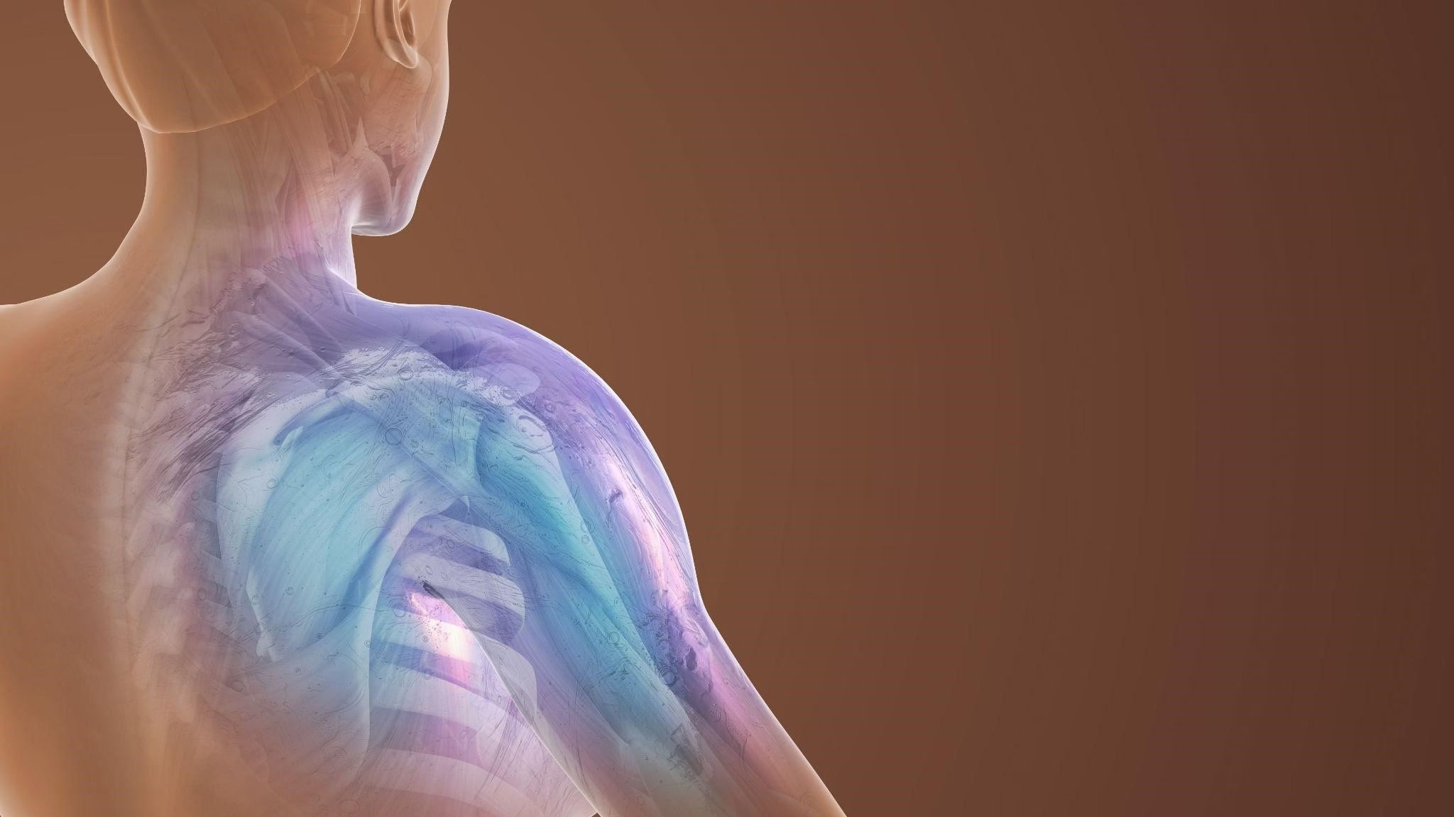
The shoulder is a ball-and-socket joint surrounded by a flexible capsule that allows a wide range of motion.

In frozen shoulder, this capsule becomes inflamed, thickened, and tight. Scar tissue gradually forms inside the joint capsule, reducing its elasticity and restricting movement.

As the capsule tightens, every shoulder movement becomes increasingly painful and limited.

Who Is Most at Risk?

Frozen shoulder can affect anyone but is more common in:

  • Adults between 40 and 65 years old
  • Women
  • People with diabetes
  • Individuals with thyroid disorders
  • Patients who have undergone shoulder surgery
  • People who have kept the shoulder immobilized after a fracture or injury
  • Patients recovering from prolonged illness

Sometimes, no obvious cause can be identified.

Common Symptoms

Symptoms usually appear gradually and worsen over time.

Patients often experience:

  • Persistent shoulder pain
  • Difficulty lifting the arm
  • Pain during the night
  • Loss of shoulder flexibility
  • Difficulty reaching behind the back
  • Pain while dressing or grooming
  • Difficulty driving
  • Stiffness that affects both active and passive movement

Unlike rotator cuff injuries, even when someone else tries to move your shoulder, the joint remains stiff.

The Three Stages of Frozen Shoulder

Stage 1 – Freezing Phase

This initial stage usually lasts several weeks to several months.

During this period:

  • Pain gradually increases.
  • Shoulder movement begins to decrease.
  • Night pain becomes common.
  • Daily activities become more difficult.

Stage 2 – Frozen Phase

Pain often becomes less intense, but stiffness is at its worst.

Patients may struggle with:

  • Reaching overhead
  • Washing their hair
  • Fastening a seatbelt
  • Reaching behind their back
  • Putting on clothes

This stage can last several months.

Stage 3 – Thawing Phase

During the final stage, shoulder mobility slowly improves.

Pain continues to decrease, and normal function gradually returns.

Recovery can take several months, and patience is important.

How Is Frozen Shoulder Diagnosed?

Diagnosis begins with a detailed medical history and physical examination.

Your orthopedic surgeon evaluates:

  • Active range of motion
  • Passive range of motion
  • Muscle strength
  • Shoulder stability
  • Areas of tenderness

A key finding is that both active and passive movements are restricted, helping distinguish frozen shoulder from other shoulder conditions.

Imaging

X-rays are often performed to rule out arthritis or fractures.

Ultrasound or MRI may be recommended to evaluate the rotator cuff and exclude other causes of shoulder pain.

Conditions That Can Mimic Frozen Shoulder

Several conditions may produce similar symptoms, including:

  • Rotator cuff tears
  • Shoulder impingement syndrome
  • Calcific tendinitis
  • Shoulder osteoarthritis
  • Cervical spine disorders
  • Shoulder instability

A careful examination helps establish the correct diagnosis.

Non-Surgical Treatment

Most patients improve without surgery.

Activity Modification

Avoid painful overhead activities while maintaining gentle daily movement to prevent further stiffness.

Medication

Anti-inflammatory medications may help reduce pain during the early stages, allowing more comfortable participation in rehabilitation.

Physical Therapy

Physical therapy is one of the most important treatments.

A structured rehabilitation program focuses on:

  • Gentle stretching
  • Restoring shoulder mobility
  • Improving flexibility
  • Strengthening surrounding muscles
  • Gradually returning to normal function

Exercises should be progressive and performed without forcing excessive pain.

Corticosteroid Injections

Ultrasound-guided corticosteroid injections can significantly reduce inflammation and pain, particularly during the early “freezing” stage.

Pain relief often allows patients to participate more effectively in physical therapy.

Hydrodilatation

Hydrodilatation involves injecting sterile fluid into the shoulder joint under imaging guidance.

The goal is to gently stretch the tight joint capsule, reduce stiffness, and improve movement.

For selected patients, this can provide excellent results when combined with rehabilitation.

When Is Surgery Needed?

Surgery is considered only when symptoms persist despite several months of appropriate conservative treatment.

Patients may be candidates if they continue to experience:

  • Severe stiffness
  • Persistent pain
  • Significant functional limitation
  • Failure to improve with physical therapy and injections

Arthroscopic Capsular Release

Arthroscopic capsular release is a minimally invasive procedure performed through small incisions.

Using a tiny camera and specialized instruments, the surgeon carefully releases the contracted capsule, allowing the shoulder to regain its normal range of motion.

The procedure typically results in:

  • Reduced pain
  • Improved mobility
  • Faster rehabilitation
  • Small scars
  • Earlier return to daily activities

Physical therapy begins soon after surgery to maintain the restored movement.

Recovery

Recovery varies from patient to patient.

Many individuals improve significantly with conservative treatment, while surgical patients often notice progressive improvement over the following weeks and months.

Following your rehabilitation program consistently is essential for achieving the best outcome.

Can Frozen Shoulder Be Prevented?

Although not all cases can be prevented, the risk may be reduced by:

  • Beginning shoulder motion early after injury when medically appropriate
  • Following rehabilitation programs after surgery
  • Managing diabetes effectively
  • Avoiding prolonged shoulder immobilization
  • Seeking early medical evaluation when pain and stiffness develop

Frequently Asked Questions

Can frozen shoulder heal on its own?

Many patients eventually improve, but recovery may take one to three years. Early treatment often shortens recovery and improves function.

Is frozen shoulder the same as a rotator cuff tear?

No. A rotator cuff tear primarily affects muscle and tendon function, whereas frozen shoulder is caused by tightening and inflammation of the joint capsule.

Is surgery always necessary?

No. The majority of patients improve with conservative treatment, including physical therapy, medications, and injections.

Can frozen shoulder come back?

Recurrence in the same shoulder is uncommon, but some people may later develop frozen shoulder in the opposite shoulder.

When should I see an orthopedic surgeon?

If shoulder pain and stiffness continue to worsen, interfere with sleep, or limit everyday activities, an orthopedic evaluation is recommended. Early diagnosis allows treatment to begin before stiffness becomes severe.

Conclusion

Frozen shoulder can be frustrating, but it is a highly manageable condition. With an accurate diagnosis, appropriate rehabilitation, and timely treatment, most patients regain excellent shoulder function and return to their normal activities.

Whether treatment involves physical therapy, image-guided injections, or minimally invasive arthroscopic surgery, the goal is always the same: relieve pain, restore movement, and help you get back to living comfortably.

Schedule a Consultation

If you are experiencing persistent shoulder pain, stiffness, or difficulty moving your arm, early evaluation can make a significant difference.

Dr. Jean-Jacques Yaacoub specializes in the diagnosis and treatment of shoulder conditions, offering individualized care ranging from conservative management to advanced arthroscopic surgery when indicated.

Book your consultation today to begin your journey toward a pain-free and more mobile shoulder.

Achilles Tendinitis: Treating Heel Pain Before It Becomes Chronic

Understanding Achilles Tendinitis

The Achilles tendon is the strongest tendon in the body, yet it is frequently injured due to repetitive stress and overload.

Achilles tendinitis commonly affects runners, athletes, active individuals, and even patients with prolonged standing activities.

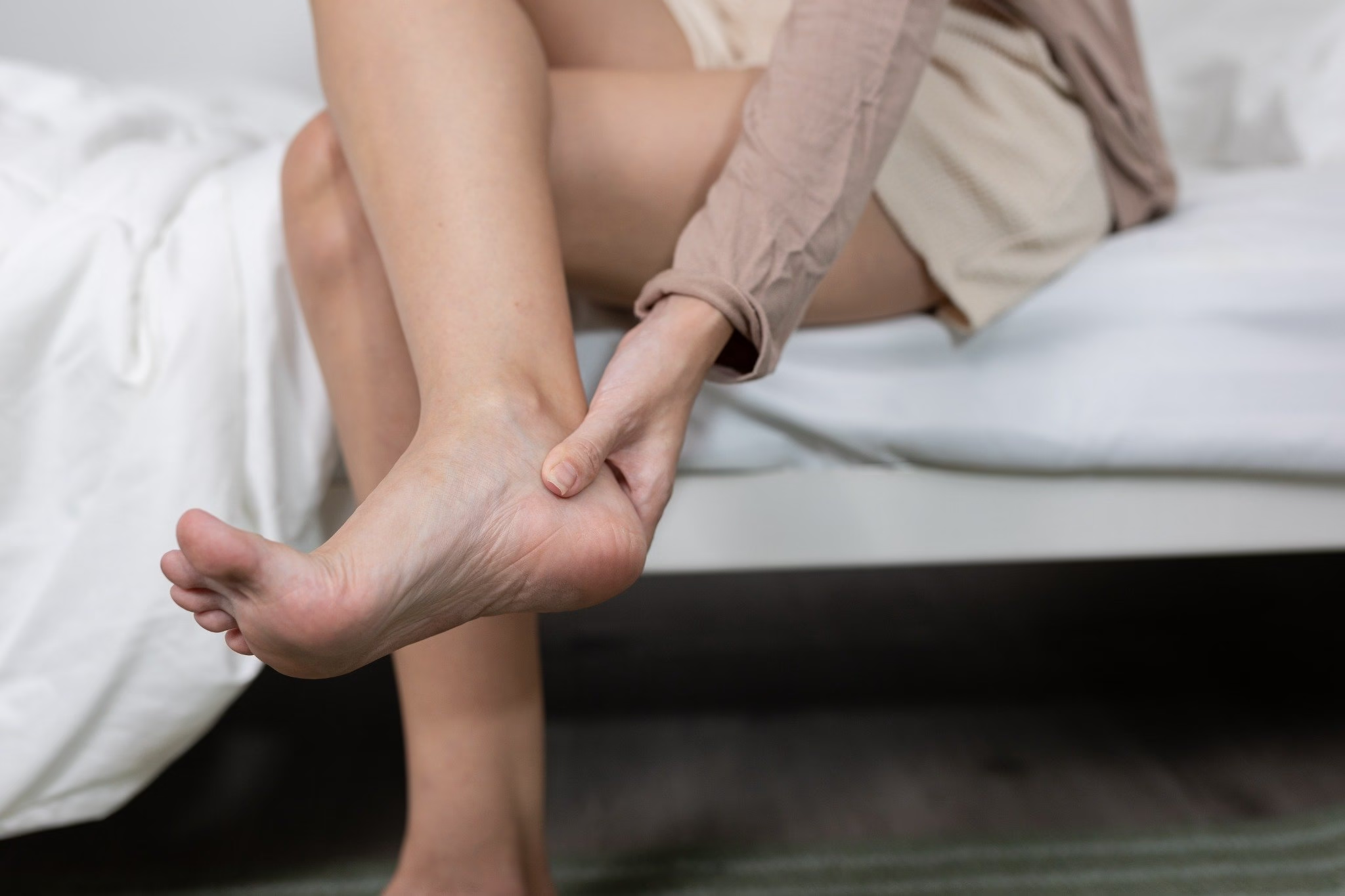
Pain at the back of the ankle may begin progressively and worsen with walking, sports, climbing stairs, or morning activity.

If left untreated, the condition can become chronic and significantly affect mobility and quality of life.

Common Symptoms

Patients with Achilles tendinitis often describe:

  • Pain behind the ankle
  • Morning stiffness
  • Pain during sports or walking
  • Tenderness along the tendon
  • Swelling or thickening of the tendon
  • Tight calf muscles

Ultrasound examination is often very helpful to evaluate tendon thickening, inflammation, microtears, or degenerative changes.

My Philosophy of Treatment

The Achilles tendon requires a precise and progressive treatment strategy.

My philosophy is based on avoiding unnecessary surgery whenever possible while treating both the tendon and the biomechanical factors contributing to overload.

Rehabilitation and Dry Needling

One of the key aspects of treatment is addressing calf muscle tightness and lower limb mechanics.

Dry needling may help reduce muscular tension in the calf muscles and improve flexibility and tendon loading patterns.

This is often combined with:

  • Eccentric strengthening exercises
  • Stretching protocols
  • Activity modification
  • Footwear optimization
  • Progressive rehabilitation

Ultrasound-Guided Infiltration

In selected chronic cases, ultrasound-guided procedures may be considered.

Precision is extremely important around the Achilles tendon because certain injections directly inside the tendon may increase rupture risk. Ultrasound guidance improves safety and accuracy.

Depending on the condition, regenerative treatments such as PRP may also be discussed.

When Is Surgery Necessary?

Surgery is considered only for resistant chronic cases, tendon tears, or major degenerative changes that fail conservative management.

Surgical treatment may involve:

  • Removal of degenerative tissue
  • Tendon repair
  • Tendon reinforcement procedures

However, many patients recover successfully through structured non-surgical treatment.

A Modern and Personalized Orthopedic Approach

Successful Achilles tendinitis treatment requires more than simply reducing pain.

My approach combines:

  • Precise diagnosis
  • Functional assessment
  • Ultrasound evaluation
  • Dry needling
  • Rehabilitation
  • Image-guided infiltrations
  • Surgery only when truly necessary

The goal is to restore movement, function, and long-term tendon health while helping patients safely return to their daily and athletic activities.

Tennis Elbow: Why Elbow Pain Is Not Always About Tennis

What Is Tennis Elbow?

Tennis elbow, also known as lateral epicondylitis, is one of the most common causes of elbow pain. Despite its name, most patients who suffer from tennis elbow are not tennis players.

This condition results from degeneration and overload of the extensor tendons of the forearm, particularly where they attach to the outer side of the elbow.a

Repetitive movements, lifting activities, sports, manual work, prolonged computer use, and overuse can all contribute to tendon irritation.

Symptoms of Tennis Elbow

Typical symptoms include:

  • Pain on the outer side of the elbow
  • Pain when lifting objects
  • Weak grip strength
  • Pain during twisting movements
  • Difficulty performing repetitive hand activities
  • Forearm tightness and muscular overload

In chronic cases, the tendon may become degenerative rather than simply inflamed.

My Treatment Philosophy

In my practice, treatment focuses on identifying the true source of pain and restoring tendon function progressively.

Many patients are told they need surgery too early, while in reality most cases improve with a structured conservative approach.

The Role of Dry Needling

Dry needling is particularly useful in patients with associated muscular tension in the forearm.

The forearm muscles often become overloaded and develop trigger points that increase stress on the tendon insertion. Treating these muscular imbalances may significantly reduce pain and improve recovery.

Dry needling is frequently combined with rehabilitation exercises to optimize tendon healing.

Ultrasound-Guided Infiltration and PRP

When symptoms persist despite physical therapy, infiltration treatments may be considered.

Corticosteroid Infiltration

Corticosteroid injections may provide short-term pain relief in highly inflammatory phases. Ultrasound guidance improves precision and safety.

PRP Infiltration

For chronic degenerative tendinopathy, Platelet-Rich Plasma (PRP) may stimulate biological healing of the damaged tendon tissue.

PRP is especially useful in resistant chronic cases where tendon degeneration is present.

When Is Surgery Considered?

Surgery is only indicated after failure of prolonged conservative treatment.

The surgical procedure generally involves removing degenerative tendon tissue and stimulating healing.

Fortunately, the majority of patients improve without surgery when treatment is personalized and progressive.

Treating the Cause, Not Only the Symptoms

My approach combines:

  • Detailed clinical evaluation
  • Ultrasound assessment when needed
  • Rehabilitation
  • Dry needling
  • Ultrasound-guided infiltrations
  • Surgery only as a final option

The objective is to help patients return to daily activities, sports, and work safely while avoiding unnecessary invasive treatment whenever possible.

De Quervain’s Disease: When Thumb Pain Starts Limiting Your Life

Understanding De Quervain’s Tenosynovitis

Pain at the base of the thumb can make simple daily activities surprisingly difficult. Opening a jar, lifting a child, typing on a phone, or even turning a key may become painful. One of the most common causes of this pain is De Quervain’s tenosynovitis.

This condition occurs when the tendons responsible for thumb movement become inflamed as they pass through a narrow tunnel on the wrist side of the thumb. Repetitive hand use, overuse, sports, lifting activities, and hormonal changes can all contribute to this painful condition.

Common Symptoms

Patients with De Quervain’s disease often experience:

  • Pain on the thumb side of the wrist
  • Pain when gripping or lifting objects
  • Swelling near the thumb base
  • Difficulty using the hand during daily activities
  • Sharp pain when moving the thumb
  • Weakness during pinching movements

The diagnosis is usually confirmed clinically during examination and may be supported with ultrasound imaging to evaluate tendon inflammation.

My Treatment Philosophy

At my practice, the goal is always to start with the least invasive and most effective solution possible.

Many patients improve significantly without surgery when the condition is treated early and precisely.

Conservative Treatment First

Initial treatment may include:

  • Activity modification
  • Wrist and thumb support
  • Anti-inflammatory treatment
  • Physical therapy
  • Dry needling for associated muscular tension

Dry needling can help reduce muscular overload around the forearm and improve tendon mechanics by decreasing abnormal tension patterns contributing to inflammation.

Ultrasound-Guided Infiltration

When pain persists despite conservative care, an ultrasound-guided corticosteroid infiltration may provide excellent relief.

Using ultrasound guidance allows precise placement of the medication around the inflamed tendon sheath while minimizing risks to surrounding structures. Precision matters, especially in delicate areas such as the wrist where skin and tendons are superficial.

Many patients experience rapid pain relief and regain normal hand function after infiltration.

When Is Surgery Necessary?

Surgery is reserved for resistant cases that fail conservative management and infiltrations.

The procedure consists of releasing the tight tendon compartment to allow the tendons to glide normally again. It is generally a short and effective surgery with good outcomes when appropriately indicated.

However, surgery should not be the first step. Proper diagnosis, rehabilitation, and targeted treatment often avoid unnecessary procedures.

A Personalized Approach

Every patient is different. My philosophy is based on combining accurate diagnosis, modern minimally invasive techniques, rehabilitation, and surgery only when truly necessary.

The objective is not simply to reduce pain temporarily, but to restore long-term hand function and quality of life.

Meniscus Tear Surgery in Dubai: When Is an Operation Truly Necessary?

Learn when meniscus tear surgery in Dubai is necessary, which tears require surgery, and when
conservative treatment is no longer sufficient.

Meniscus Surgery Is About Timing, Not Urgency
Avoiding surgery at all costs is not the goal.
Rushing into surgery without proper indication is equally problematic.

The real question is:
When does surgery become the best solution for this knee?

Symptoms That May Require Meniscus Surgery
Meniscus surgery in Dubai is considered when patients experience:
 True mechanical locking of the knee
 Recurrent catching or blocking
 Persistent pain despite proper conservative treatment
 Recurrent knee effusions
 Functional limitation affecting work or daily life
These signs suggest the tear is mechanically unstable, not just visible on MRI.

Meniscus Tears That Often Require Surgery
Surgery is more commonly indicated in:
 Bucket-handle tears
 Displaced meniscal fragments
 Acute traumatic tears
 Meniscus tears associated with ACL injuries
 Tears causing repeated mechanical symptoms
In these cases, injections and physiotherapy alone are unlikely to resolve the problem.

Surgical Options for Meniscus Tears in Dubai
The surgical strategy depends on:
 Tear type and location
 Vascular supply of the meniscus
 Patient age and activity level
Meniscal Repair (Suture)
 Preferred whenever possible
 Preserves meniscal function
 Requires structured rehabilitation

 Best for young or active patients
Partial Meniscectomy
 Removal of unstable fragments only
 Aims to relieve mechanical symptoms
 Should always be as conservative as possible
Total meniscectomy should be avoided due to long-term cartilage damage.

Surgery Is Only Part of the Treatment
Successful meniscus surgery depends on:
 Proper indication
 Precise surgical technique
 Structured postoperative physiotherapy
 Gradual return to activity
Surgery without rehabilitation is incomplete treatment.

Long-Term Objective
The goal of meniscus surgery is not only pain relief,
but long-term knee preservation.

If knee pain persists despite conservative treatment,
or if you experience locking or instability,
a specialist meniscus surgery evaluation in Dubai can determine whether surgery is truly
necessary and which option best preserves your knee.

Meniscus Tear Treatment in Dubai: Why Conservative Care Comes First

Meniscus tear treatment in Dubai using a conservative approach. Learn when injections and
physiotherapy can relieve knee pain without surgery.

Knee Pain and Meniscus Tears: A Common Situation in Dubai
In Dubai, many patients consult for knee pain after an MRI report shows a meniscus tear. Very
often, they are advised surgery immediately, even when symptoms are moderate.
However, a meniscus tear on MRI does not automatically explain knee pain, and it certainly
does not always require surgery.
The correct approach starts with understanding the clinical behavior of the knee, not just
imaging results.

Treating the Knee, Not the MRI
MRI technology is extremely sensitive. In adults, especially after the age of 35, meniscus lesions
are frequently found in pain-free knees.
In clinical practice:
 Pain often comes from inflammation, overload, or muscle imbalance
 The meniscus tear may be present but not mechanically problematic
 Operating based on imaging alone can accelerate cartilage damage
This is why conservative treatment is often the first-line option for meniscus tears in Dubai.

When Conservative Meniscus Treatment Is Appropriate
A non-surgical approach is usually indicated when:
 Knee pain developed progressively
 There is no true locking of the knee
 Full knee extension is preserved
 Swelling is intermittent

 Pain worsens with activity but improves with rest
These situations represent the majority of meniscus consultations in Dubai.

Conservative Meniscus Tear Treatment in Dubai: My Philosophy
Conservative care is not passive care.
It is a structured strategy combining injection therapy and physiotherapy.

Ultrasound-Guided Knee Injections
When pain or inflammation limits movement, physiotherapy alone often fails.
In these cases, ultrasound-guided knee injections are used to:
 Reduce intra-articular inflammation
 Control pain precisely
 Create the conditions needed for effective rehabilitation
Injections are:
 Image-guided for accuracy
 Used to support recovery, not to mask pain
 Always combined with rehabilitation
An injection without physiotherapy is incomplete treatment.

Physiotherapy for Meniscus Tears
Physiotherapy is the cornerstone of conservative treatment.
Rehabilitation focuses on:
 Quadriceps and hamstring strengthening
 Hip and gluteal muscle control
 Proprioception and knee stability
 Correction of movement patterns
 Load management for daily activities and sports
A meniscus does not fail in isolation.

It suffers when knee biomechanics are altered.

Expected Results With Conservative Meniscus Treatment
When correctly indicated, patients often experience:
 Progressive pain reduction
 Improved knee confidence
 Decreased swelling
 Return to daily activities
 Avoidance of surgery in many cases
Most importantly, this approach preserves the meniscus and protects long-term knee health.

Key Principle
A meniscus tear does not automatically require surgery.
Conservative treatment should always be considered first.

If you were advised knee surgery based mainly on MRI findings,
a personalized meniscus evaluation in Dubai can determine whether conservative treatment
with injections and physiotherapy is still appropriate.

Why Does My Shoulder Hurt?

Shoulder pain is one of the most common orthopedic complaints. It can range from mild discomfort — like difficulty brushing your hair — to intense, stabbing pain that wakes you at night.

But what causes shoulder pain? And what’s really going on inside the joint?

Understanding the Shoulder Joint
The shoulder is a complex structure made of bones, tendons, ligaments, and bursae. Its main function is to allow a wide range of movement. So when movement becomes painful, what’s usually to blame?

The answer is often subacromial impingement.

What Is Subacromial Impingement?
Subacromial impingement happens when the rotator cuff tendons rub against the acromion (a bony projection of the scapula). This repeated friction causes inflammation of the tendons (tendinitis) and the nearby bursa (bursitis), resulting in pain, especially when lifting the arm.

Standard Treatment vs. Advanced Options
Traditional treatment includes NSAIDs and physiotherapy. While this helps some patients, others may not improve for weeks or even months.

In our practice, we offer a faster and more precise solution using ultrasound-guided infiltration.

What Is Ultrasound-Guided Infiltration?
Here’s how the procedure works:

  • A topical anesthetic is applied to numb the skin.
  • Under real-time ultrasound, we insert a needle directly into the inflamed bursa.
  • A targeted injection — either corticosteroids or PRP (Platelet-Rich Plasma) — is delivered precisely where it’s needed.

This reduces pain and inflammation almost immediately, allowing faster return to movement and improved outcomes with physical therapy. Many patients feel better within a day.

Why Ultrasound Matters
Unlike blind injections, ultrasound ensures that medication reaches the exact area of inflammation. If you’ve had a cortisone shot that didn’t work, it may not have been placed correctly.

Precision matters. That’s why we use ultrasound for every injection we perform.

When Is Surgery Needed?
In more advanced cases, especially with persistent or recurrent symptoms, we may consider arthroscopic subacromial decompression. This minimally invasive surgery removes bone spurs and reduces pressure on the rotator cuff tendons, allowing proper movement to return.

Final Thoughts
Most cases of shoulder pain can be treated without surgery. The key is early diagnosis, precise imaging, and the right intervention.

Stay tuned for future posts where I’ll cover more causes and treatments for shoulder pain.

Why Your Shoulder Pain Keeps Coming Back

Shoulder pain that returns after every round of physical therapy or cortisone injections can be frustrating and concerning. Many patients come to our clinic saying, “I thought I was better, but the pain came back after a few weeks.”

So, why does this happen?

The Root Cause Was Never Fully Treated

Pain relief is only one part of healing. In many cases, the inflammation subsides, but the structural issue — like a tendon tear, calcium deposit, or impingement — was never fully resolved. This leads to repeated flare-ups.

Common culprits:

  • Partial rotator cuff tears.
  • Undiagnosed calcific tendinitis.
  • Subacromial bursitis from ongoing impingement.


The Injection Missed the Target

Many cortisone or PRP injections are done “blind” — without imaging. If the medication doesn’t reach the exact area of inflammation, the relief is incomplete and short-lived.

In my clinic, every injection is performed under real-time ultrasound guidance to ensure precision. We don’t guess—we treat what we see.

You’re Stronger… But Not Stabilized

Pain might go away after rest or therapy, but if the rotator cuff and scapular muscles aren’t properly reconditioned, the shoulder remains vulnerable. A small movement or poor posture is enough to cause a setback.

That’s why we work closely with physical therapists to build strength and stability after pain relief.

What You Can Do About It

  • Get a precise diagnosis using ultrasound or MRI.
  • Treat inflammation and the underlying structural issue.
  • Follow a personalized rehab plan that focuses on long-term function.
  • Don’t settle for temporary relief — aim for complete recovery.

Shoulder pain doesn’t have to be part of your routine. We use a combination of modern diagnostics and targeted treatments to help patients heal smarter and stay pain-free.